Blood biomarkers

What a blood test can — and cannot — tell you

NeuroGlympse offers blood biomarker testing alongside objective functional assessment — because the two answer different questions, and a brain injury is characterised properly only when you have both.

The four markers

What each protein actually reports

Different proteins leak from different cells on different timelines. That is why the timing of a blood draw changes what you can learn from it.

GFAP

Glial fibrillary acidic protein

Released from
Astrocytes
Detection window
Rises within hours, peaks around 24 hours

A structural protein of astrocytes. Its appearance in blood indicates astroglial injury, and it is one of the two markers in the FDA-cleared tests used to decide whether a head CT is needed.

UCH-L1

Ubiquitin C-terminal hydrolase L1

Released from
Neuron cell bodies
Detection window
Rises fast, clears fast — within the first day

An enzyme abundant in neuronal cell bodies. Paired with GFAP because the two together are more informative than either alone; its short half-life makes early sampling essential.

S100B

S100 calcium-binding protein B

Released from
Astrocytes (and tissues outside the brain)
Detection window
Peaks around day 1, near-normal by days 2–12

The longest-established marker, used in European guidelines to help avoid unnecessary CT. Its weakness is specificity: it is also released by fat, muscle and skin, so orthopaedic injury or hard physical exertion can raise it without any brain injury at all.

NF-L

Neurofilament light chain

Released from
Long myelinated axons
Detection window
Rises later and stays up for months

A scaffolding protein of the neuronal cytoskeleton, concentrated in subcortical white-matter axons. In one serial study plasma NF-L was roughly 483× baseline at day 2, 89× at day 9, and still 3× at day 176 — which makes it a marker of axonal damage over time rather than a snapshot.

Three different questions

Biomarkers, imaging and function are not substitutes

Each modality answers something the others cannot. Treating any one of them as the whole answer is how injuries get missed.

Comparison of what blood biomarkers, CT and MRI imaging, and functional ocular motor testing each measure after a head injury
ModalityQuestion it answersUseful windowBlind spot
Blood biomarkersHas brain tissue been damaged biologically?Hours to days (NF-L: months)Cannot tell you how the brain is functioning
CT / MRIIs there bleeding or a structural lesion?AcuteUsually normal in mild TBI
Symptom questionnaireWhat does the patient report feeling?Any timeSelf-reported, and easily masked or over-reported
Ocular motor testingIs brain function measurably impaired?Best within 3 weeksSignal fades as the brain compensates

Where NeuroGlympse fits

After the scan comes back clean

The patient who benefits most from understanding biomarkers is the one whose results were normal. The emergency department did its job: it asked whether there was bleeding, and the answer was no.

What nobody has measured yet is whether the brain is working properly. That is a functional question, and it needs a functional test — which is why our assessment measures ocular motor performance and then follows autonomic recovery through continuous monitoring.

Biomarkers and functional testing are complementary, not competing. One tells you tissue was injured; the other tells you what that injury is doing to the person.

We offer both, which is the point — a biomarker result read next to an ocular motor result is far more informative than either read alone.

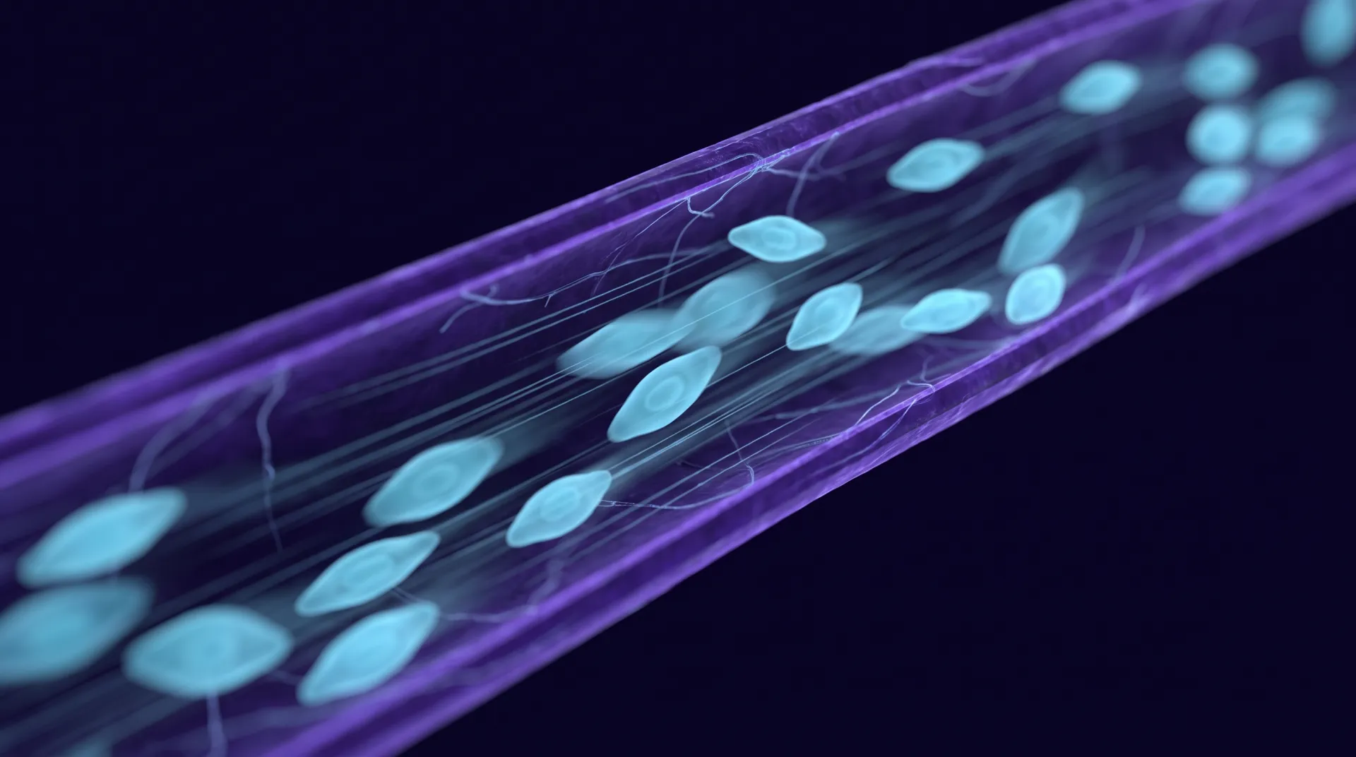
Macro abstraction of microvascular blood flow, individual cells rendered as soft luminous forms moving through a vessel.
Blood-based markers reveal that tissue was injured. They cannot report how the injury is affecting function.

The direction of travel

No single test defines a brain injury

In 2025 the NIH-NINDS TBI Classification and Nomenclature Initiative proposed the CBI-M framework: characterising a traumatic brain injury across four axes — Clinical findings, Biomarkers, Imaging, and Modifiers such as prior injury and comorbidity — instead of collapsing it into a single severity label.

It is a formal acknowledgement of what clinicians treating these patients already knew: mild, moderate and severe were never adequate, and no one modality is sufficient on its own.

Clinical

Symptoms, examination findings and functional performance — including ocular motor testing.

Biomarker

Blood proteins reporting cellular and axonal injury.

Imaging

CT and MRI, for structural damage.

Modifiers

Prior injuries, comorbidities and the factors that shape recovery.

Common questions

Blood biomarkers, answered plainly

Does NeuroGlympse offer blood biomarker testing?

Yes. We provide biomarker testing alongside our functional ocular motor assessment, with results interpreted by our board-certified neurology team and read together rather than in isolation. Contact our team and we will advise which panel is appropriate for how long ago the injury happened.

Is there a blood test that diagnoses a concussion?

No. This is the single most misunderstood point. The FDA-cleared blood tests measuring GFAP and UCH-L1 are cleared to help decide whether a patient needs a head CT — that is, to rule out bleeding or a structural lesion. A negative result is good news about your skull; it is not a statement that your brain function is unaffected. Concussion remains a functional diagnosis.

Which blood tests are actually FDA cleared?

Abbott's i-STAT TBI test measuring GFAP and UCH-L1 was cleared in 2021 for plasma, with a whole-blood point-of-care cartridge cleared in 2024. bioMérieux's VIDAS TBI, measuring the same two proteins, was cleared in 2024. An earlier assay from Banyan Biomarkers was first cleared in 2018. All are indicated for evaluating whether imaging is needed after mild TBI.

How accurate are they?

For their actual indication — detecting intracranial injury visible on CT — the reported performance is strong: roughly 96.7% sensitivity and a 99.4% negative predictive value, with results in about 18 minutes. That high negative predictive value is the point: the test is very good at telling clinicians who does not need a scan.

If my blood test was normal, why do I still have symptoms?

Because the test was answering a different question. Most mild traumatic brain injuries produce no bleeding and no structural lesion, so both the CT and the biomarker panel come back reassuring while the functional injury — the disrupted eye movement, the disturbed sleep, the suppressed heart rate variability — is entirely real and measurable by other means.

How soon does blood need to be drawn?

Very early for GFAP and UCH-L1, which rise and clear within roughly the first day; the FDA-cleared indications are built around acute presentation. S100B peaks around day 1 and returns near baseline within a couple of weeks. NF-L is the exception — it climbs later and stays elevated for months, so it is the one marker still informative well after the event.

What is the CBI-M framework?

A 2025 framework from the NIH-NINDS TBI Classification and Nomenclature Initiative proposing that TBI be characterised across four axes rather than a single severity label: Clinical findings, Biomarkers, Imaging, and Modifiers such as prior injury and comorbidity. It is a formal acknowledgement that no one test defines a brain injury.

Our testing

Biomarker testing through NeuroGlympse

We provide blood biomarker testing as part of the same assessment pathway as our functional testing, so a single referral produces both the biological and the functional picture rather than sending your patient to two unconnected providers.

Results are interpreted by our board-certified neurology team and read alongside ocular motor findings and monitoring data — not returned as an isolated number for someone else to make sense of.

One referral, both axes

Biomarker draw and functional assessment ordered together, with a single point of contact for the result.

Neurologist-interpreted

A clinician reads the panel in the context of the injury, the timeline and the functional findings.

Carried into monitoring

Where a marker suggests ongoing axonal injury, remote therapeutic monitoring follows the recovery objectively rather than by symptom recall.

Order both, and read them together

Biomarker testing and objective functional assessment from one referral, interpreted by the same neurology team.

Call usEnroll a patient