
TMS · the science
Changing a circuit, not just the chemistry
Transcranial magnetic stimulation treats depression by inducing electrical activity in a specific cortical target and driving lasting plasticity in the network it belongs to. Here is the mechanism, in detail.
Why a different mechanism matters
Antidepressants and TMS do not do the same thing
Most antidepressants act on neurotransmitter availability — raising synaptic serotonin, norepinephrine or dopamine and waiting for downstream adaptation. That works for a great many people. When it does not, prescribing a fourth drug with the same basic mechanism has diminishing returns, which is the defining frustration of treatment-resistant depression.
TMS intervenes at a different level. Rather than altering chemistry diffusely across the brain, it delivers energy to a specific cortical location and changes the excitability and connectivity of the circuit that location belongs to. That is why failing medication does not predict failing TMS: the two are not competing attempts at the same intervention.
Step 1 — the physics
A magnetic pulse becomes an electric current inside the brain
An electromagnetic coil sits against the scalp. A capacitor discharges through it in well under a millisecond, producing a magnetic pulse of roughly 1.5 to 2 tesla — comparable to an MRI scanner, but lasting microseconds instead of minutes.
The reason this is non-invasive comes down to physics: bone and skin are effectively transparent to a magnetic field, whereas they scatter and attenuate electrical current badly. So the field crosses the skull essentially undiminished, and by Faraday’s law of induction, a rapidly changing magnetic field induces an electric current in any nearby conductor — including cortical tissue. Where that induced current is strong enough, neurons depolarise and fire.
The field weakens sharply with distance, which is the single most important constraint on the whole therapy: TMS can only directly stimulate the outer couple of centimetres of cortex. Everything deeper has to be reached indirectly.
Step 2 — the target
Stimulating what you can reach to change what you cannot
The therapeutic target in depression sits deep in the brain. The solution is to stimulate a surface region that is wired to it.
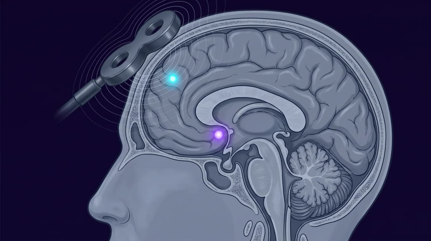
Left DLPFC
Directly stimulated. The field reaches only the outer couple of centimetres of cortex — watch the contours fade well before the midline.
Subgenual cingulate
The therapeutic target, below the front curve of the corpus callosum. No coil reaches it; it responds through its connection with the DLPFC.
The conventional target is the left dorsolateral prefrontal cortex, which is characteristically underactive in depression and happens to lie within reach of a coil. But the more interesting part of the story is what it connects to.
The left DLPFC is functionally anticorrelated with the subgenual anterior cingulate cortex — when one is more active, the other tends to be less so. The subgenual cingulate is heavily implicated in depression and sits far too deep for any coil. Research indicates that the strength of that anticorrelation at the stimulation site is associated with how well a patient responds,3 which is why connectivity-guided targeting has become a serious area of work rather than simply measuring five centimetres forward from the motor cortex.
Recent intracranial recordings in humans have shown directly that stimulating the DLPFC evokes responses in the subgenual cingulate,4 and work on white-matter routes between the two suggests the length of that pathway helps explain who responds.5 The clinical implication is straightforward: where the coil is placed matters as much as how much energy it delivers.
Step 3 — the dose
Frequency decides whether you excite or suppress
Before any treatment, your individual motor threshold is measured — the minimum intensity that produces a small twitch when the coil is over the motor cortex. Treatment intensity is then set as a percentage of that threshold, so dosing is calibrated to your own anatomy rather than a fixed machine setting. Consensus guidance is to recheck it during the course.
Frequency determines direction. Stimulation at around 10 Hz and above tends to increase cortical excitability, which is why high-frequency stimulation is applied to the underactive left DLPFC. Stimulation at about 1 Hz tends to decrease it, which is why low-frequency protocols are sometimes applied to the right side instead — the same logic, approached from the opposite direction.
High frequency, left
Low frequency, right
Theta burst
Step 4 — the plasticity
Why the effect accumulates instead of wearing off
A single session shifts cortical excitability briefly and then fades. The antidepressant effect depends on something more durable: long-term potentiation-like strengthening of synapses in the stimulated circuit — the same class of activity-dependent plasticity the brain uses for learning. Repeated, patterned stimulation is what converts a transient change into a structural one.
This is the honest explanation for the treatment schedule. A full course is roughly 30 to 36 sessions, typically five days a week for four to six weeks, with most patients noticing change around weeks two to three. Improvement that lags the first sessions is not the treatment failing; it is what plasticity accumulating looks like.
Pulse
The induced current depolarises neurons in the target region, forcing synchronous activity.
Train
Patterned trains within a session repeatedly co-activate connected neurons — the precondition for synaptic strengthening.
Course
Across sessions, those changes consolidate. Excitability and connectivity in the network shift measurably.
Circuit
Signalling between prefrontal cortex, cingulate and salience-network regions changes direction, and mood follows.
Theta burst
Three minutes doing the work of thirty-seven
Intermittent theta burst stimulation delivers very short bursts — triplets at about 50 Hz — repeated at roughly 5 Hz, the frequency of the brain’s own theta rhythm. Patterning stimulation on an endogenous rhythm induces plasticity considerably more efficiently than uniform pulse trains, so far less time is needed for the same effect.
The THREE-D trial tested this properly: 414 patients with treatment-resistant depression randomised to conventional 10 Hz stimulation lasting about 37.5 minutes, or iTBS lasting about 3 minutes. iTBS was non-inferior. Roughly 50% of patients responded and about a third reached remission.1 The FDA cleared iTBS for major depressive disorder in 2018.
Accelerated protocols push further. Stanford’s SAINT/SNT approach uses functional-connectivity-guided targeting and delivers ten sessions a day for five days, reporting high remission rates in a treatment-resistant cohort without cognitive side effects.2 It received FDA clearance in 2022. Availability is still limited and it requires imaging-based targeting, but it demonstrates how much of the outcome depends on where and how densely you stimulate.
The evidence
What the numbers actually show
414
patients randomised in THREE-D, comparing 3-minute iTBS with 37.5-minute 10 Hz stimulation1
~50%
responded to iTBS in THREE-D — in patients who had already failed medication1
~33%
reached remission in THREE-D, with iTBS non-inferior to conventional rTMS1
3 min
iTBS session length versus 37.5 minutes, for equivalent outcome1
These are the randomised trial figures. Registry and real-world series report higher response rates than medication trials in comparable populations, but they span different protocols, populations and remission definitions, so we do not quote a headline number for them here. What matters is that THREE-D enrolled patients for whom medication had already failed.
Safety
What the risks genuinely are
Common and transient
- Scalp discomfort or tapping sensation during stimulation
- Headache after a session, usually easing over the first week
- Facial muscle twitching while the coil is active
- No anaesthesia, no sedation, no recovery period
Rare and serious
Seizure is the meaningful risk. Under consensus screening and dosing it runs at roughly 1 per 30,000 sessions or lower, with reported rates around 0.31 per 10,000. Every reported TMS seizure has occurred during stimulation, been self-limiting, and left no lasting sequelae.6
Ferromagnetic metal in or near the head is the principal contraindication. Screening precedes any stimulation.
Indications
Who it is cleared and used for
Major depressive disorder
Treatment-resistant depression
Everything else is off-label
After a head injury
Why this sits inside a concussion care model
Major depressive disorder is reported in 33–42% of patients within the first year after a traumatic brain injury.7 Those patients are frequently already contending with cognitive symptoms, fatigue, sleep disruption and a growing medication list — which makes a non-pharmacological option that does not impair cognition unusually well matched to the situation.
Because we monitor mood, sleep and heart rate variability continuously, deterioration tends to surface in the data rather than waiting for a patient to volunteer how bad things have become at a quarterly appointment. That flags a case for clinical review. It does not select a treatment.
Practicalities
What a course actually involves
Session length
Course length
When it works
Afterwards
Questions
TMS, answered properly
How does TMS actually work?
A coil held against the scalp carries a brief, very strong current, which produces a magnetic pulse of roughly 1.5 to 2 tesla. Magnetic fields pass through skin and bone essentially unimpeded, and inside the brain that changing field induces a small electric current in cortical tissue — Faraday's law of induction. Where the induced current is strong enough it depolarises neurons and makes them fire. Repeated in patterned trains over several weeks, this drives lasting changes in synaptic strength within the circuits being stimulated.
Why the prefrontal cortex, specifically?
The left dorsolateral prefrontal cortex is typically underactive in depression, and it is one of the few nodes of the depression network that a coil can physically reach — the field falls off sharply and only penetrates a couple of centimetres. Crucially, the left DLPFC is functionally connected to the subgenual anterior cingulate cortex, a deep region strongly implicated in depression that no coil can reach directly. Stimulating the accessible node modulates the deeper one through that connection.
Why does it take weeks rather than one session?
A single session produces a transient change in cortical excitability that fades. The therapeutic effect depends on plasticity — long-term potentiation-like strengthening of synapses — which accumulates only with repeated, patterned stimulation. That is why the consensus is a full course of roughly 30 to 36 sessions, and why stopping early tends to waste the treatment rather than deliver a smaller version of it.
What is theta burst stimulation, and is it as good?
Intermittent theta burst stimulation delivers short 50 Hz triplets repeated at about 5 Hz — a pattern that mirrors the brain's own theta rhythm and induces plasticity far more efficiently. The THREE-D trial randomised 414 patients with treatment-resistant depression to standard 10 Hz stimulation lasting about 37.5 minutes versus iTBS lasting about 3 minutes, and iTBS was non-inferior (Blumberger et al., THREE-D, Lancet 2018). The FDA cleared iTBS for major depressive disorder in 2018.
How effective is it?
In the THREE-D trial roughly 50% of patients responded and about a third reached remission, in a population where medication had already failed (Blumberger et al., Lancet 2018). Registry and real-world series generally report higher response than medication trials in comparable patients, but they span different protocols, populations and remission definitions, so we do not quote a single headline figure for them. For treatment-resistant depression these are meaningful numbers, because each successive medication trial tends to deliver less.
How safe is it? Is it like ECT?
No — it is a genuinely different procedure. There is no anaesthesia, no induced seizure, and no memory impairment; patients drive themselves home and return to normal activity immediately. The main risks are scalp discomfort and headache during or shortly after a session, both usually transient and easing over the first week. Seizure is the serious risk and it is rare: under consensus screening and dosing, roughly 1 per 30,000 sessions or lower, with reported rates around 0.31 per 10,000 (Brain Stimulation, survey of over half a million sessions, 2021). Every reported TMS seizure has occurred during stimulation, been self-limiting, and left no lasting sequelae.
Who should not have TMS?
Ferromagnetic metal in or near the head — certain aneurysm clips, cochlear implants, some stimulator hardware — is the principal contraindication, because the magnetic field interacts with it. A history of seizures, or medications and conditions that lower seizure threshold, calls for careful assessment rather than automatic exclusion. Screening happens before any stimulation and includes measuring your individual motor threshold.
Why consider TMS after a concussion?
Major depressive disorder is reported in 33–42% of patients within the first year after a traumatic brain injury (J Neuropsychiatry Clin Neurosci, systematic review and meta-analysis). Those patients are often already managing cognitive symptoms, fatigue and a medication burden, which makes a non-pharmacological option that causes no cognitive impairment particularly relevant. Because we monitor mood and physiology continuously, candidacy tends to surface from the data rather than waiting for someone to raise it at an appointment.
References
Every figure on this page traces to a source below, so the claims can be checked rather than taken on trust.
- 1.Blumberger DM, et al. (THREE-D trial). Effectiveness of theta burst versus high-frequency repetitive transcranial magnetic stimulation in patients with depression (THREE-D): a randomised non-inferiority trial. The Lancet. 2018;391(10131):1683–1692.
- 2.Cole EJ, et al.. Stanford Accelerated Intelligent Neuromodulation Therapy for treatment-resistant depression. American Journal of Psychiatry. 2020;177(8):716–726.
- 3.Review. Mechanisms of action of TMS in the treatment of depression. Current Topics in Behavioral Neurosciences. 2024.
- 4.Human intracranial EEG study. Dorsolateral prefrontal cortex TMS evokes responses in the subgenual anterior cingulate cortex. bioRxiv / PMC.
- 5.Imaging study. White matter pathways mediating dorsolateral prefrontal TMS therapy for depression. Nature Neuroscience.
- 6.Survey of over half a million treatment sessions. Seizure risk with repetitive TMS. Brain Stimulation. 2021.
- 7.Systematic review and meta-analysis. Predictors of major depression and post-traumatic stress disorder following traumatic brain injury. Journal of Neuropsychiatry and Clinical Neurosciences.
This page is educational and is not medical advice. Prevalence figures are presented as the source reports them, including confidence intervals and ranges; a single number would imply more precision than the evidence supports.
Find out whether TMS is appropriate for you
Candidacy is assessed by our clinical team, taking your treatment history, screening findings and monitoring data into account.